|


|

IM512P Data and
Image Processor
 
 
-
There are two models of procesor: IM512P and
IM512P/E. They differ basically in two features of
acquisition software:
v IM512P/E corrects energy,
linearity and uniformity in real time when used to upgrade gamma cameras;
v IM512P/E's
acquisition zoom values are more flexible and fully
digital.
-
Processing software is exactly the same for
both models.
-
When the IM512P/E is required, it is mentioned with its full name (for
instance, when describing the upgrades for gamma cameras). The generic
mentions of IM512P refer to both models.
-
The IM512P may be connected to any
gamma camera with analogic X,Y & Z output. In this general kind of cameras,
the IM512P/E only makes use of the uniformity correction and more
flexible and accurate acquisition zoom.
-
The IM512P/E makes a full use of its
abilities in the upgrade of GE Starcam, Siemens Orbiter or Elscint Apex gamma
cameras. Details of these upgrades may be seen by clicking on Gamma Cameras
button of this page and then looking at the respective presentations.
-
The IM512P may be used also as an
only-processing work station with gamma cameras that keep their original
acquisition computer. At present, there are IM512Ps working in
communication with the following models of divers manufacturers:
▪
New!
ADAC Pegasys: network transfer in native format, format conversion and
insertion into IM512P data base. Fully automatic, studies arrive as they are
acquired. Compatible with modality worklist (MWL).
▪ GE Millennium MG, MPR and MPS, and Optima NX [DICOM
transfer].
▪ Siemens ICON, e.soft and c.Cam [DICOM transfer].
▪ Sopha/SMV DS7 Acquisition Station [FTP transfer in native format].
▪ Elscint XPert [FTP transfer in Interfile format].
▪ Philips Cardio MD acquisition computer [DICOM transfer].
▪ Picker
Prism 1000
[FTP transfer in Interfile format].
▪ Picker
Odyssey FX-800
[DICOM transfer].
Acquisition
-
No restriction for
simultaneous acquisition and processing. You may be acquiring a static,
dynamic, planar gated, whole body, SPECT
or gated
SPECT study and processing any other planar or SPECT study at the same time.
-
Double 14 bits AD
converter (for X and Y axes). Optional 14
bits converter for energy.
-
Acquisition matrices: 1024 x 1024, 512 x 512, 256 x 256, 128 x 128
and 64 x 64, up to
255 or 65.535 counts per pixel.
-
Double isotope acquisition with simultaneous viewing
of both images in different windows. Up to 3 energy windows into 1, 2 or 3
simultaneous images. (IM512P/E)
-
Dynamic acquisition: more than 5000 64x64
images. Up to 1000 images per second (1 ms/image).
-
X and Y
COR correction "on the fly".
Theoretical accuracy:
1/4000 FOV (0.1 mm).
-
Built-in ECG for cardiac
studies, with its cable set.
-
Programmable acquisition presets.
-
Persistence image
with real time
zoom x 1; x 1.5; x 2; x 3; x 4 and x 6. Variable acquisition X-Y
displacement to bring any point of the image into the acquisition matrix
center by clicking on it. Optional (IM512P/E):
zoom x 1.25; x 1.75; x 2.5; x 3.5; x 5; x 7
and x 1.33.
-
Simultaneous viewing of ECG, cine display and images during gated
acquisition.
-
Automatic saving of images during
acquisition.Minimum loss in case of blackout.
-
On the fly energy,
linearity and uniformity correction. Each pair isotope/collimator
may have its own correctio map. 1024 channel pulse height analyzer. Double isotope/triple
window with energy correction. It replaces the gamma camera console. (IM512P/E)
Screen
Display
-
Configurable
display (minimum: 1024 x 768) that allows seeing on the same screen:
▪
Several studies in different windows simultaneously.
▪
One or more text
editors to write study reports.
-
The
mouse helps to move,
change size and arrange images to print.
-
Each image window may
have its own color table, upper and lower levels to get the optimal
brightness and contrast
for each one.
-
More than 200 color tables may be created by
users.
-
64k simultaneous colors
on screen, selected from a palette of 262,144 colores. Programmable colors
for regions of interest,
curves, characters, markers, etc.
-
More than 1000 regions
of interest per study (rectangular, circular, elliptical, irregular,
by gradient).
-
More than 1000
histograms or curves per study.
-
More then
16 simultaneous cine
displays on screen, each one with its own variable rate and zoom.
-
Screen may be copied to Windows*
clipboard
and pasted to any graphic application.
Files
-
Hard disk storage of
studies with patient data, images, ROIs,
curves, annotations, etc. (more than de 1 million 64 x 64 images).
-
DVD drive. CD, pen
drive or external hard disk may be used also. Option TCP/IP, NetBEUI,
DICOM servers.
-
Patient/study data
base that allows recording of oral reports.
Planar Clinical Protocols
-
Ejection
fraction,
peak and average ejection rate, peak filling rate, time to peak filling rate, Fourier
phase analysis, wall motion.
Quantification of
Myocardial Perfusion with Interpolated Background Subtraction and
Circumferential Profiles. Shunt detection. First Pass.
-
Protocols for Renal Perfusion, Renogram, Renal Quantitation,
GFR with DTPA (with and without blood sample), ERPF with MAG3 and one blood
sample, Brain Flow, Esophageal Transit, Gastroesophageal Reflux, Pulmonary
Quantification, Field Uniformity quality control, Gall Bladder Ejection
Fraction, Gastric Emptying, Parathyroid (Tl-Tc subtraction), Thyroid Uptake,
etc.
-
A whole
body can be reconstructed from a series of spots.
-
Several
studies can be processed simultaneously in different windows.
-
Commands
for general use to process images, regions of interest and curves.
-
Recording
and reproduction of sequenced orders to automate jobs.
-
High
processing speed: 0.9 seconds (CPU time) for an Ejection Fraction while
another dynamic or gated study is acquired.
SPECT Clinical
Protocols (optional)
-
Tomographic Reconstruction with multiple filters, movement and attenuation
correction at 0.01 seconds (CPU time) per slice
-
Tridimensional Analysis, 3D Surface Maps, X, Y, Z Axes Reorientation (0.004
sec. per reoriented slice). Interpolated Zoom. Stress and Rest Gated SPECT.
-
Real
time reslicing.
-
Gated
Polar Map, final display in 5 seconds.
-
Image
fusion with other modalities.
-
Summed,
Maximum Activity and Weighted Transparency reprojections.
-
3D volume
viewer that shows six slices on the six faces of a 3D perspective cube that
can be rotated, moved and resized with the mouse
Gated SPECT
(optional)
-
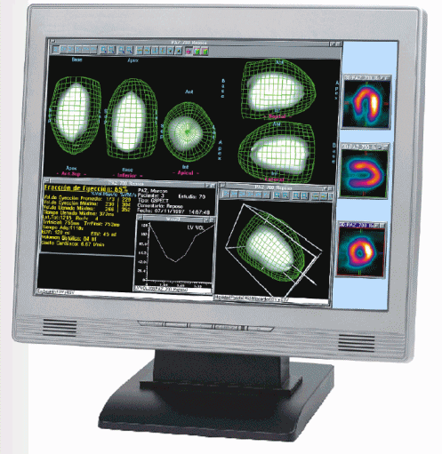
Volume
calculation protocol from gated SPECT images (SPECT sync with ECG) begins
with an automatic reorientation of the cardiac axes and automatic search of
the limits of the left ventricle (LV).
-
The
method is based on fitting a truncated ellipsoid to the actual volume of the
LV. Then endo- and epicardial edges are detected along 829 radii
perpendicular to the ellipsoid surface, for every interval in the cardiac
cycle, constrained to the fact that ventricular mass must remain constant
through the cycle. The fitted ellipsoid is used only as a starting point in
order to generate the profiles. Each profile is analyzed in search of the
endocardium, the epicardium and the midmyocardium (a point of maximal uptake
in the LV wall), by using several methods: maximum of the filtered profile
or most probable value in a fitted Gaussian curve for midmyocardium,
inflexions or weighted standard deviation in a fitted Gaussian curve for
endo- and epicardium. The detected points (one for each profile) generate a
final contour that is not the original fitted ellipsoid but the actual LV
surface. All the process takes only a few seconds with current PC
processors.
-
Endocardial detected positions allow the calculation of the end diastole
(ED) and end systole (ES) volumes and from there the ejection fraction is
calculated, global or at each point of the ventricle surface (regional
ejection fraction). Wall thickness and thickening, and LV mass are also
estimated.
-
Endo-,
epi- and midmyocardium can be shown at ED and ES, or beating along the
cardiac cycle. Both situations allow the appreciation of the wall motion.
-
The
reconstructed ventricular volume can be used to map the perfusion, the
absolute thickness, the thickening, the wall motion or the ejection
fraction, in colors. This way, in each cine 3D image the shape, the motion
and the functional parameter can be evaluated in colors.
-
This
protocol can show simultaneously these results from 5 points of view:
anterosuperior, posteroinferior, septal, lateral and apex. Or in a
perspective volume that the operator moves at will, interactively with the
mouse.
-
This
protocol can show simultaneously up to four studies of one or different
patients.
-
This
allows the operator to see in cine five stress 3D surfaces plus other five
of rest and also for pre and post treatment. (Total 20 cine surfaces).
-
This
protocol generates volumes, positions and thicknesses for each of the radii
and cardiac intervals in a table compatible with Excel* or similar, for
future analysis. For quality control it is possible to plot, total or
partially, the 829 curves (profiles) of each interval of the cardiac cycle.
-
The
analysis displays allow to display as a solid surface the epi-, mid- or
endocardium and as a mesh surface (wire mesh) the epi- or midmyocardium, to
appreciate two simultaneous surfaces in cine mode and from any angle, by
rotating the images with the mouse.
-
Also the
traditional slices of short axis and horizontal and vertical long axis can
be generated, automatically reoriented and realigned between stress and
rest, cine or static. Summed polar
maps can be generated. Also eight cardiac interval polar maps can be
created, displaying the change of activity between ED and ES in cine mode.
-
Each
display with its settings may be stored for later review with a single click
in Next Screen / Next Study.
Documentation
-
Color
printer with photographic quality in sheets of 8.5” x 11”, with up to 6
photos per sheet.
-
DICOM
printer.
-
Laser
printer, black and white or color
-
Ink-jet
printer
-
Any
printer for Windows* can be used
-
.JPG, .PNG or
.PCX files for image interchange;
.AVI or .WMV for movies.
-
Transmission of studies through local network or Internet without stopping the acquisition
or processing.
-
Screen copy and paste
to other Windows* applications.
Acquisition
from several Gamma Cameras (optional)
-
Simultaneous acquisition from two gamma cameras with only one PC, each with
its respective ECG trace, with simultaneous display of all acquisitions.
This acquisition also allows simultaneous processing
-
Simultaneous acquisition from multiple gamma cameras with one PC for each
camera, connected by Ethernet. Each PC can be an acquisition, or a
processing or an acquisition and processing workstation
DICOM
Connectivity
(optional)
_____________________
*
Windows and Excel are registered trademarks of Microsoft Corp.
Other brand or product names are trademarks or registered trademarks of their respective
holders.
This software has
the clearance 510K of the Food and Drug Administration (FDA).
Copyright
Alfanuclear S.A.I. y C., 1994-2015
 |